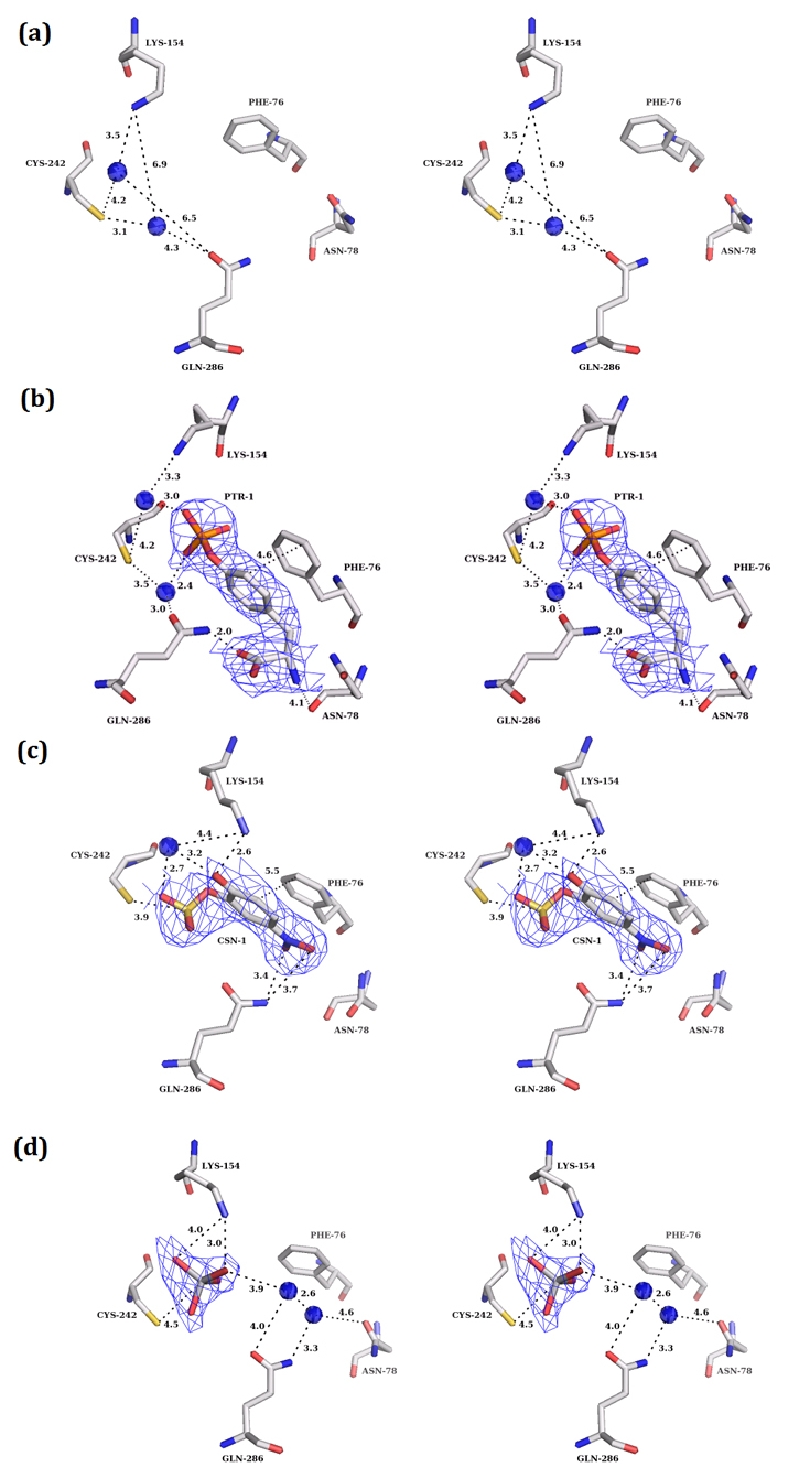
Figure 3. The apo and the substrate/inhibitor bound active site of PTP10D.

(mFo-DFc) electron density maps (at 3.0σ level) for the apo and substrate/inhibitor bound states of PTP10D. a. The active site of apo PTP10D (PDB: 3S3E; resolution 2.4Å). b. The phosphotyrosine of the GP4 peptide at the active site of PTP10D (PDB ID: 3S3H; resolution 2.8Å). c. PNC bound at the active site of PTP10D (PDB ID: 3S3K; resolution 3.2Å). d. Vanadate bound at the active site of PTP10D (PDB ID: 3S3F; 2.7Å).
