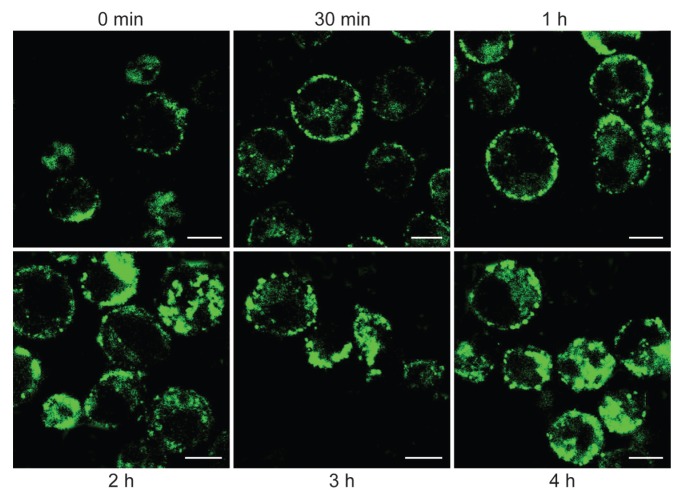
Figure 1.
Confocal microscopy analysis of CD38 internalization. DL06 myeloma cells were incubated with saturating amounts of FITC-labeled agonistic IB4 mAb for 30 min at 4°C. After rinsing in RPMI-1640 medium (Sigma, Milano, Italy) with 5% fetal calf serum (Euroclone, Milano, Italy), affinity-purified goat anti-mouse IgG2a (SouthernBiotech, Birmingham, AL, USA) was added for 10 min at 4°C. Cells were successively incubated at 37°C for selected times. DL06 were then fixed at 4°C for 20 min in PBS containing 4% formalin and rinsed in PBS. The samples were analyzed with an Olympus FV300 laser scanning confocal microscope equipped with a Blue Argon (488 nm) laser and FluoView 300 software (Olympus Biosystems, Hamburg, Germany).

