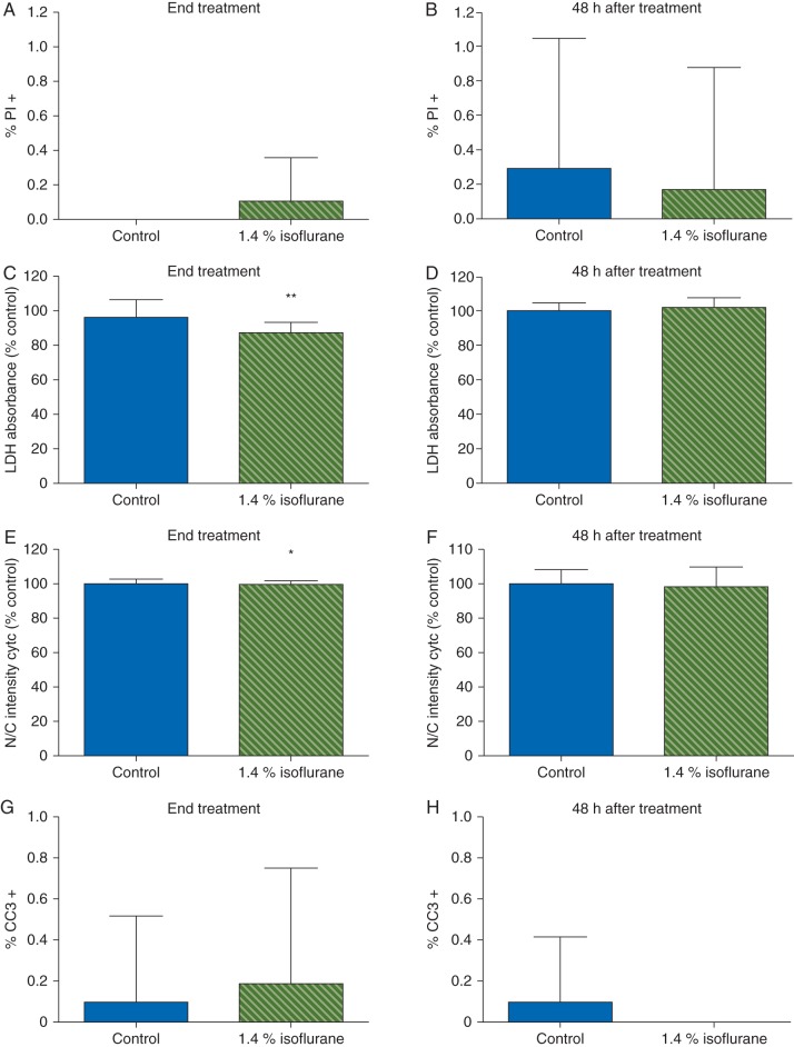
Fig 4.
Isoflurane did not alter astrocyte viability. Astrocytes were exposed to control conditions or 1.4% isoflurane for 4 h. (a, b) Very few cells stained positive for PI under control conditions and isoflurane had no effect on the percentage of astrocytes stained by PI at the end of exposure 48 h later. (c) Isoflurane decreased LDH release in astrocytes during exposure but there were no effects on LDH activity 48 h later (d). There was a small but statistically significant decrease in nuclear translocation of cytochrome C during isoflurane exposure (e) but no effect (f) 48 h later. Similarly, there were very few control or isoflurane-treated cells that stained positive for cleaved caspase 3 either at the end of exposure (g) or 48 h later (h). Taken together these data suggest that isoflurane prevents endogenous astrocyte death during exposure but has no effect on necrotic cell death 48 h later. Data expressed as mean (sd), n=12–20 wells per treatment condition, *P<0.05, **P<0.001.

