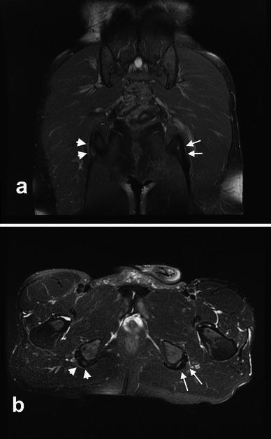
Fig. 5.

MRI of a 30 years old male long-distance runner 18 months after surgical treatment for left PHT. a Proton density-weighted coronal image showing no abnormal signal intensity of the left proximal hamstring insertion (arrows). The right proximal hamstring tendons present increased signal intensity compatible with PHT (arrowheads). b T2-weighted axial image clear shows no intratendinous structural abnormalities in the left proximal hamstring tendons (arrows). The right side presents signs of PHT (arrowheads). This patient underwent surgical treatment for right PHT four months after having performed the MRI, but he was not considered in this study because of the follow-up inferior to 24 months
