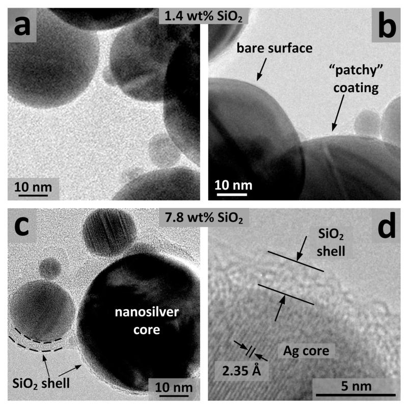
Figure 3.
TEM images of the 1.4 wt% (a,b) and 7.8 wt% (c,d) SiO2 coated nanosilver. Patchy coatings of nanosilver with bare surface as well as with very thin (<1 nm), non-continuous amorphous SiO2-coating are observable (b). The nanosilver core can be distinguished from its amorphous silica coating (c). The distance of the crystal planes (ca. 2.35 Å) corresponds to the (111) plane of Ag metal (d).

