
Figure 1.
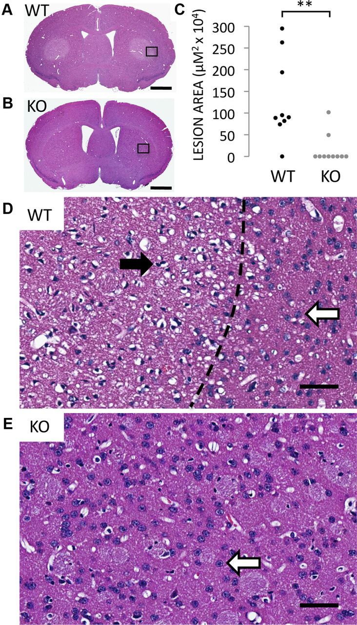
Rhes-deleted mice are protected from striatal lesions caused by systemic 3-NP intoxication. A, B, H&E stain of striatum from WT (A) and Rhes-deleted (KO) mice (B) treated with 3-NP. Scale bar, 1 mm in A and B. C, Area of striatal lesions caused by 3-NP; n = 9 for WTs, n = 10 for KOs; **p < 0.005. D, High-magnification view of region indicated in A. Dotted line indicates border between lesioned and normal striata. Closed arrowhead points to a pyknotic nucleus from an apoptotic neuron, and open arrowheads point to normal nuclei from healthy neurons. E, High-magnification view of region indicated in B. Scale bars, 50 μm for D and E.