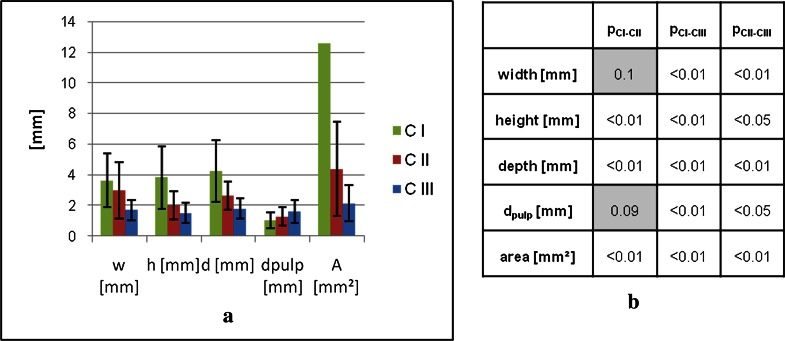
Figure 4.
(a) Geometrical properties of the lesions in the different categories obtained by the three-dimensional ultrashort echo time (3D UTE) technique. Figure 1b shows the significance of the differences. The standard deviation (±14.2 mm) for the CI area is omitted in the visualization. CI, class 1; CII, class 2; CIII, class 3; dpulp, minimal distance between lesion and pulp.

