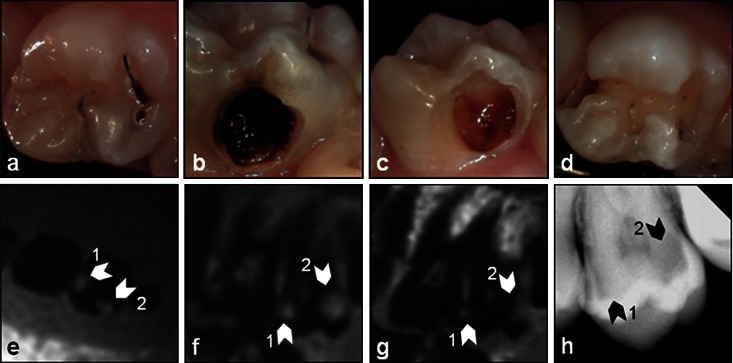
Figure 5.
Smooth surface caries (CI): intraoral images (a, occlusal; b, bucal) prior to the dental treatment and the images (c, bucal; d, occlusal) after excavation of the lesions and the respective (e) transversal and (f) parasagittal three-dimensional ultrashort echo time (g) multislice turbo spin-echo and (h) X-ray images of the tooth. The white arrows in the MR images and the black arrows in the X-ray image indicate the lesions

