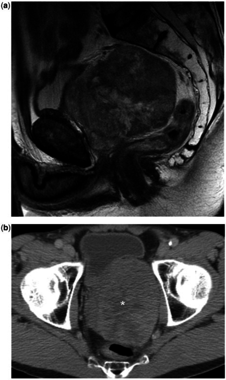
Figure 6.
A 54-year-old man with a prostatic leiomyosarcoma. (A) Sagittal T2-weighted MR image shows the tumour as a large, well-defined, rounded mass arising from the superior aspect of the prostate, with heterogeneous T2 signal intensity. (b) Axial CT image shows the lesion as a heterogeneous mass, containing areas of necrosis/cystic change (asterisk).

