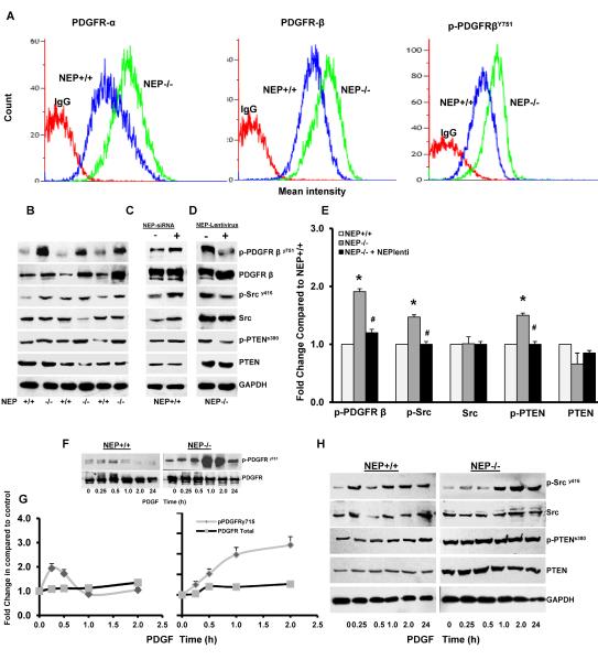
Figure 3. Loss of NEP increases PDGFR expression and signaling in NEP−/− PASMCs.
Flow cytometry analysis was performed on NEP+/+ and NEP−/− PASMCs to assess levels of PDGFRα, β and p-PDGFR751 expression. An overlay from a representative paired isolate is shown in Panel A. Lysates were analyzed for levels of phospho- and total PDGFR, Src and PTEN at baseline in NEP+/+ and NEP−/− PASMCs by Western blot shown in Panel B. Effects of NEP siRNA (10nMole/L) in NEP+/+ PASMCs, and NEP lentivirus in NEP−/− PASMCs on expression of phospho- and total PDGFR, Src and PTEN are shown in Panels C and D, respectively. Panel E shows the fold change in expression levels of the different kinases in NEP−/− PASMCs compared to matched NEP+/+ cells for 6 different paired isolates. Data was normalized to GAPDH used as a loading control. Panel F shows time dependent changes (0–24h) in levels of p-PDGFRy715 and total PDGFR levels in response to PDGF(10ng/ml) in NEP+/+ and NEP−/− cells and Panel G a comparison plot of change with time. Panel H shows change in levels of p-Src and p-PTEN in response to PDGF (10ng/ml) in NEP+/+ and NEP−/− cells. *p≤ 0.05 for comparison between NEP+/+ and −/− PASMC. # p≤ 0.05 for comparisons between NEP−/− with and without lentivirus treatment.

