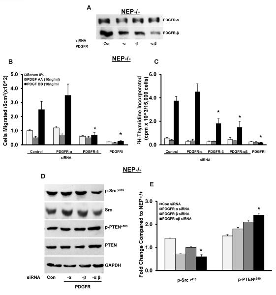
Figure 4. Knockdown of PDGFR attenuates migration and proliferation and normalizes PDGFR signaling in NEP−/− PASMCs.
PASMCs were treated with either control siRNA or siRNA to PDGFRα, β or αβ and migration and proliferation were measured after 48h. Panel A shows levels of PDGFRα and –β receptors in the presence of siRNA. PDGF AA (10 ng/ml; ligand specific for PDGFRα) and PDGF BB (10 ng/ml; specific for PDGFR β) were used to assess the contribution of each receptor to migration and proliferation in siRNA or PDGFR kinase inhibitor III (PDGFRI, 500 nM/L for 24h) treated NEP −/− cells shown in Panels B and C from three different isolates. NEP−/− PASMCs treated with PDGFRα, β and αβ siRNA were analyzed for p-PDGFR, p-Src and p-PTEN and levels are shown in Panel D. Panel E shows average levels from 3 different isolates. *p≤ 0.05 for comparisons between control siRNA and PDGFR siRNA treatment (n=3).

