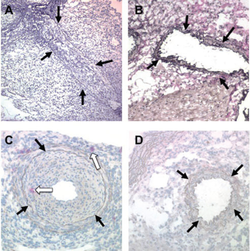
Figure 3. Coronary arteries of B10.BR heart allografts in B6.RAG1−/− complement sufficient recipients after administration of DSA with or without NK depletion.
(A) A severely stenotic coronary artery in a B10.BR allograft from a B6.RAG1−/− recipient treated with anti-H-2KkIgG2a mAb is shown, demonstrating severe CAV. (B) Addition of anti-NK1.1 mAb during anti-H-2KkIgG2a mAb administration prevents the CAV lesion. (C) Ly49g2 immunohistochemical stain of a coronary artery with CAV from a B10.BR heart in a B6.RAG1−/− recipient given anti-H-2Kk IgG2a. Red stained NK cells are present in the thickening intima and in the adventitia (white arrows). (D) Same strain combination given anti-NK1.1 in addition to anti-H-2Kk IgG2a. No Ly49g2+ cells or intimal thickening are seen. A–D, elastin stains; black arrows indicate coronary arteries

