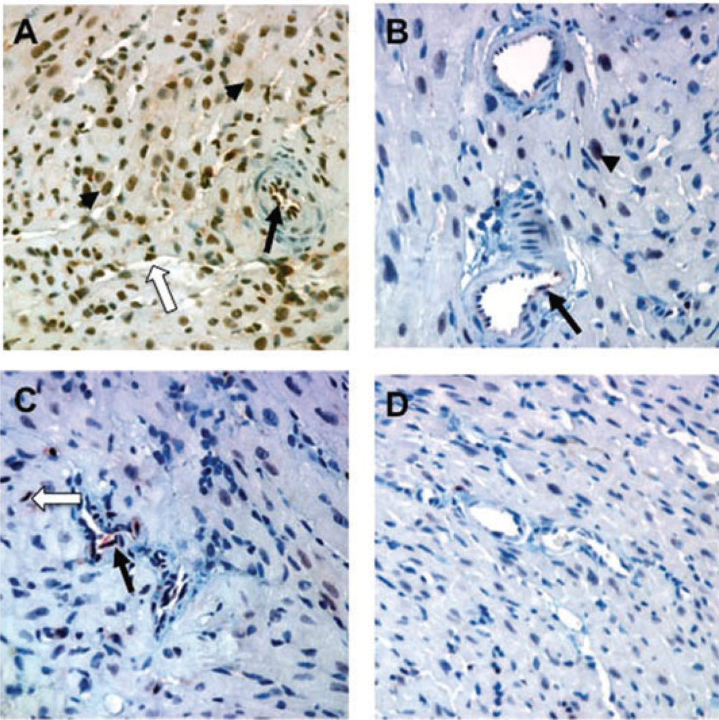
Figure 6. Effects of DSA on pERK expression in heart allograft endothelial cells on day 28.
(A) Intact anti-H-2Kk DSA (HB13) elicits a strong arterial (black arrow) and capillary (white arrow) endothelial pERK response in B10.BR allografts in B6.RAG1−/− recipients. Myocyte nuclei (black arrowhead) also showed pERK expression. (B) In contrast, little pERK was detected in B10.BR allografts in response to F(ab’)2 DSA. (C) Endothelial expression of pERK was somewhat less extensive when NK cells were depleted during the time of DSA administration. (D) Isografts in B6.RAG1−/− recipients given HB13 showed little or no increase in pERK expression. Immunhistochemical stains with anti-pERK; original magnification 400×.

