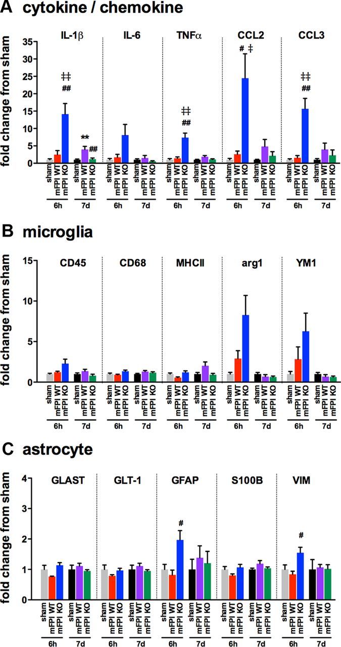
Figure 7.

Enhanced cytokine and chemokine gene expression is seen in the p38α KO mice at 6 h after diffuse brain injury. Temporal changes in cytokine/chemokine (A), microglia (B), and astrocyte markers (C) were measured from the cortex harvested from sham mice (gray represents 6 h; black, 7 d), injured WT mice (red, 6 h; purple, 7 d), or injured p38α KO mice (blue, 6 h; green, 7 d); n = 4–10 per group. **p < 0.01, sham versus mFPI WT. #p < 0.05, mFPI WT versus mFPI KO. ##p < 0.01, mFPI WT versus mFPI KO. ‡p < 0.05, sham versus mFPI KO. ‡‡p < 0.01, sham versus mFPI KO.
