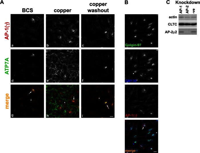
FIGURE 6:
AP-1 depletion blocks ATP7A trafficking to the TGN. (A) HeLa Cells depleted of AP-1 by transfection with siRNA targeting the μ1 subunit were treated with BCS, CuCl2, or CuCl2, followed by washout. Cells depleted of AP-1 are identified by loss of adaptin γ signal at the TGN (a–c, g–i red). Cells are counterstained for ATP7A (d–f, g–i green). Examples of depleted cells are marked in the merge panels with asterisk, and nondepleted cells are marked with an arrow. (B) AP-1–depleted cells, identified by loss of adaptin γ signal (red), are labeled for the trans-Golgi marker golgin‑97 (green) or the cis-Golgi marker GM130 (blue). Scale bar, 10 μm. (C) Immunoblot of lysates from control cells (-ve) or those depleted of AP-1 or AP-2 and blotted with antibodies indicated on the left of the blot. A total of 10 μg of protein was loaded per lane.

