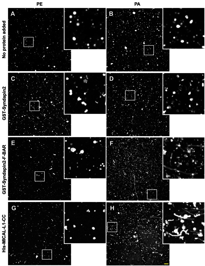
FIGURE 6:
Phosphatidic acid–rich LMVs undergo tubulation in the presence of F-BAR domain of Synd2 and CC domain of MICAL-L1. In vitro tubulation assays were performed with (A) PE-containing liposomes only, (B) PA-containing liposomes only, (C) PE-containing liposomes and GST-Synd2, (D) PA-containing liposomes and GST-syndapin, (E) PE-containing liposomes and GST-Synd2-F-BAR, (F) PA-containing liposomes and GST-Synd2-F-BAR, (G) PE-containing liposomes and His-MICAL-L1-CC, or (H) PA-containing liposomes and His-MICAL-L1-CC. LMVs comprised a mass ratio of 70% PC, 10% NBD-PE, and 20% PE or PA. Insets depict the region in the white box. Bar, 10 μm.

