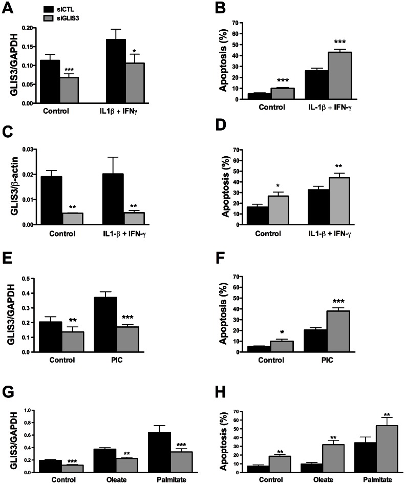
Figure 2. GLIS3 KD potentiates apoptosis induced by cytokines, PIC, and FFAs.
Following transfection with siCTL and siGLIS3 as in Figure 1, INS-1E cells (A, B, E–H) and human islet cells (C, D) were exposed to cytokines (A, B, C, D) (n = 4–7), PIC (E, F) (n = 7), oleate or palmitate (G, H) (n = 5). After 24 h GLIS3 mRNA expression and apoptosis were evaluated. Results are means ± SEM. * P<0.05, ** P<0.01 or *** P<0.001 vs. siCTL by paired t-test.

