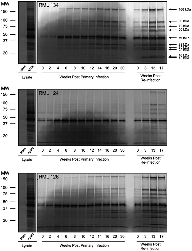
Figure 1. Antibody signature of C. trachomatis infected macaques following primary infection and re-challenge.
Three monkeys were infected ocularly with C. trachomatis strain A2497 [7]. Monkeys cleared the infection 9–14 wpi (RML134: 13 wpi; RML124: 8 wpi; RML126: 14 wpi). Clinical disease remained severe to moderately severe during the periods of culture positivity and continued until about 30 wpi. Three months after resolution of disease monkeys were re-challenged and all monkeys exhibited partial protection characterized by a more rapid clearance of organism (3–7 wpi) with an accompanying reduction in the intensity and duration of ocular inflammation (11 wpi). Sera collected from monkeys during primary infection and following re-challenge were analyzed by RIP using intrinsically labeled chlamydial proteins prepared from non-denaturing detergent lysed infected McCoy cells. Sera from all three macaques produced a similar yet simple antigen recognition profile that differed in complexity over the period of primary infection and re-challenge. A total of 10 proteins were immunoprecipitated with molecular masses of 166, 90, 72, 60, 40, 29, 25, 23, 16 and 15 kDa. There was a noticeable temporal and recall recognition pattern of the antigens in all three monkeys. The MOMP (40 kDa) recognition was strong very early post infection (2–4 weeks) and consistently sustained throughout the entire period of infection and disease. In contrast immunoprecipitation of the other chlamydial proteins occurred later following primary infection, with the strongest immunoprecipitation reactions occurring at the time of infection eradication and disease resolution. There was a recognizable increase in the immunoprecipitation of all of these proteins by all three animals following re-challenge.

