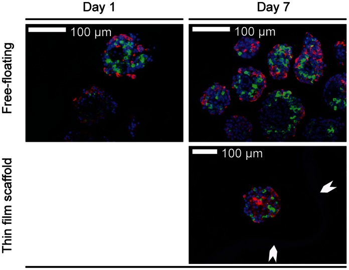
Figure 8. Histology of free-floating control islets and of islets cultured in thin film microwell scaffolds.
Insulin (red) and glucagon (green) positive cells can be observed throughout the islets. The microwell scaffold is indicated by the white arrows. All data were obtained after 7 days of culturing.

