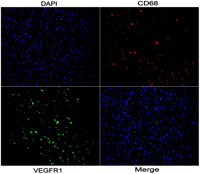
Figure 4. Location of peritumoral expression of VEGFR-1.
Co-expression of CD68 and VEGFR-1 in stromal compartments in peritumoral liver tissue. The CD68 (red) and VEGFR-1 (green) signals were due to rhodamine- and FITC-labeled antibodies, respectively, using single-layer projections in a confocal microscope. Hepatocyte nuclei were labeled by DAPI (blue).

