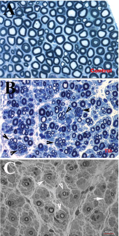
Figure 1.

(A) Sciatic nerve section from normal control autopsy, stained with Toluidine blue, contains numerous myelinated nerve fibers. (B) In contrast, a semithin section from the tibial nerve of a patient with axonal form of MPZ mutation, also stained with Toluidine blue, showed a reduced density of myelinated nerve fibers with many regenerating clusters (arrowheads). These features are consistent with axonal type of neuropathy. (C) A semithin section from a sural nerve biopsy of a patient with CMT1B showed numerous onion bulbs (arrowheads) with severely reduced densityof myelinated nervefibers. These features are typical for CMT1. (Reprinted with permission from Li J et al6 and Bai et al7)
