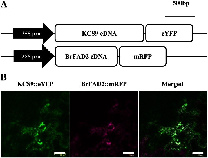
Figure 2.
Subcellular localization of Arabidopsis KCS9 in tobacco epidermis. A, Schematic diagrams of pKCS9::eYFP and pBrFAD2::mRFP constructs. 35S pro, Promoter of cauliflower mosaic virus 35S RNA. B, Fluorescent signals of KCS9::eYFP and BrFAD2::mRFP in tobacco epidermal cells. Genes encoding fluorescent proteins were translationally fused to KCS9 and BrFAD2 under the control of the CaMV 35S promoter. The constructed vectors were coinfiltrated into tobacco epidermis via A. tumefaciens-mediated transformation, and the fluorescent signals were observed using a laser confocal scanning microscope 48 h after infiltration. BrFAD2::mRFP was used as an ER marker (Jung et al., 2011). Bars = 10 µm.

