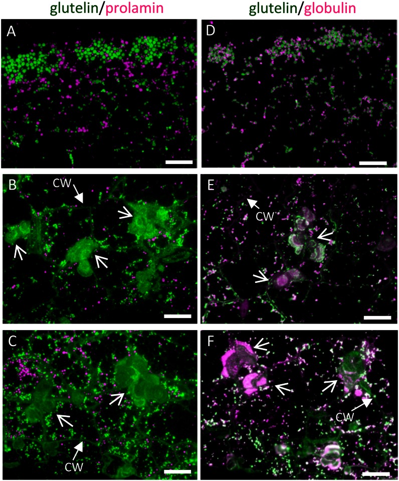
Figure 4.
The distribution of storage proteins in the wild type (A and D) and glup6 mutant lines EM939 (Gln-140 stop; B and E) and EM1327 (Gln-262 stop; C and F) as viewed by immunofluorescence microscopy. A to C depict the distribution of glutelins and prolamines, while D to F depict the distribution of glutelin and α-globulin in 3-WAF seeds. Secondary antibodies against mouse and rabbit IgGs labeled with FITC (green) and rhodamine (magenta), respectively, were used to detect antigens recognized by mouse anti-glutelin and rabbit anti-prolamine, respectively, in A to C and mouse anti-glutelin and rabbit anti-α-globulin, respectively, in D to F. Note that glutelins and globulins, which are normally packaged in PSVs (A and D), are secreted and mislocalized to the large PMBs (arrows) located adjacent to the cell wall (B, C, E, and F). CW, Cell wall. Bars = 20 μm.

