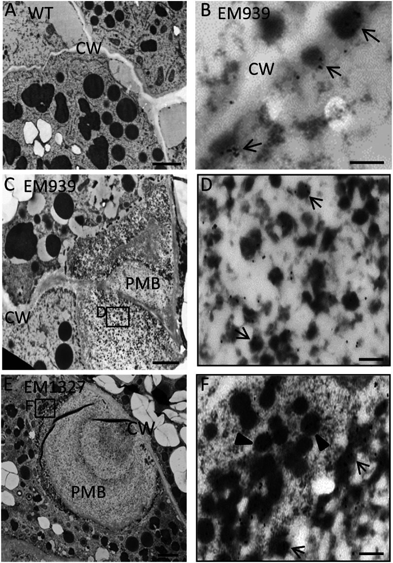
Figure 6.
Immunolocalization of glutelin to electron-dense granules and PMBs in glup6 mutants. A, The wild type (WT). B to D, EM939. E and F, EM1327. B, Seed at 1 WAF. A and C to F, Seeds at 2 WAF. D and F, Enlarged images of the areas enclosed by boxes in C and E, respectively. The secreted electron-dense granules, which are reactive to anti-glutelin (arrows), are located between the cell wall (CW) and plasma membrane (B). The granules within PMBs (D) and the dense vesicles close to the PMB in cytoplasm (F) are depicted with arrows and arrowheads, respectively. Gold particles (15 nm) indicate the reaction of anti-glutelin. Bars in A, C, and E = 2 μm; bars in B, D, and F = 200 nm.

