Figure 7.
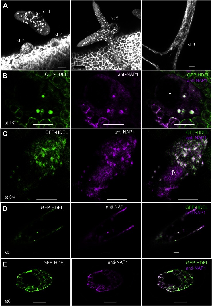
In expanding trichomes, the ER consists of a distributed population of nodule-like structures that strongly colocalize with NAP1. A, Localization patterns of GFP-HDEL in wild-type live-cell trichomes. GFP-HDEL is localized to punctate structures in early stage 2 (st 2) trichomes and slightly larger elliptical structures in stage 4 (st 4) trichomes, as shown by the maximum Z-projection of multiple confocal images. GFP-HDEL is localized to elliptical structures in stage 5 (st 5) trichomes, as shown by the maximum Z-projection of a confocal image stack containing most of the stage 5 trichome that is transitioning to a pointed branch tip morphology (middle). GFP-HDEL is localized to membrane sheets and extended tubules in stage 6 (st 6) trichomes, as shown by the maximum Z-projection of confocal images containing part of a stage 6 trichome (right). B, Single confocal image slice showing the colocalization between NAP1 (middle) and GFP-HDEL (left) in a stage 2 trichome at punctate structures, as shown by the white pixels (right). V, Lumen of the central vacuole. C, Single confocal image slice showing the colocalization between NAP1 (middle) and GFP-HDEL (left) in a stage 3/4 trichome at punctate structures, as shown by the white pixels (right). N, Nucleus. D, Single confocal image slice showing the colocalization between NAP1 (middle) and GFP-HDEL (left) in a stage 5 trichome containing punctate structures, as shown by the white pixels (right). E, Single confocal image slice showing the colocalization between NAP1 (middle) and GFP-HDEL (left) in a stage 6 trichome with a reticulate membrane organization, as shown by the white pixels (right). Bars = 10 μm in A to C and 5 μm in D and E.

