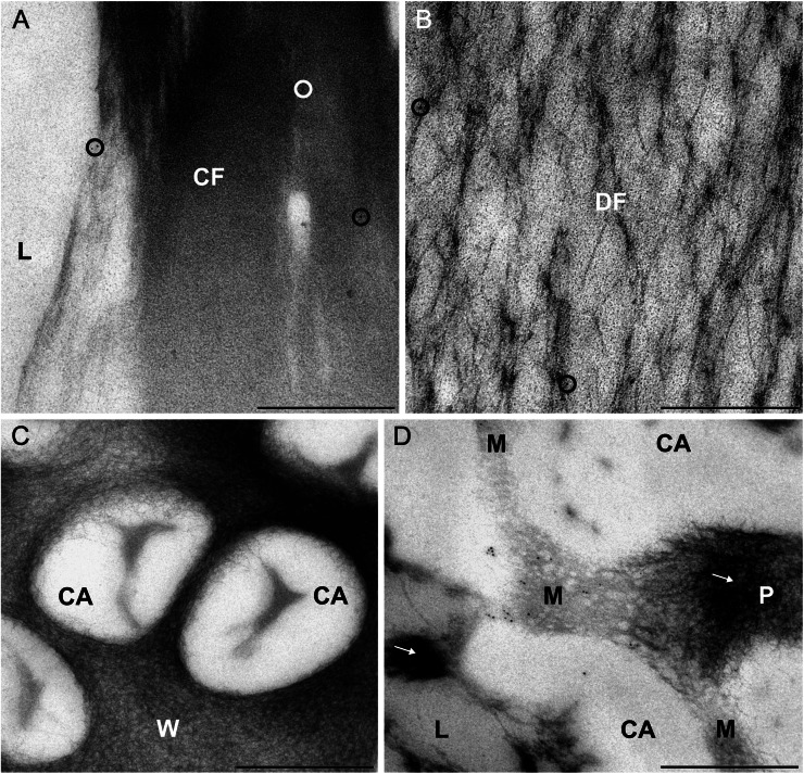
Figure 3.
Immunoreactivity of forisomes and sieve pores of fava bean SEs to actin antibodies. For indirect immunolocalization, ultrathin sections were labeled with clone C4 anti-actin (dilution, 1:200) and 5-nm gold-labeled secondary antibodies. Washing conditions were stringent. Nonspecific labeling is reduced to a negligible amount (circles in A and B) on condensed forisomes (CF in A) and dispersed forisomes (DF in B) lying in the lumen (L) of the SEs. No label occurs on the cell wall areas of the sieve plates (W in C) and on the callose collars surrounding the sieve pores (CA in C and D). A significant labeling is visible in an oblique section through a sieve pore (D), passing through the SE lumen (L) with aggregated P-proteins (arrow), the callose collar (CA), the plasma membrane and the mictoplasm (M), and the sieve pore lumen (P) plugged with P-proteins (arrow). The labeling occurs in the tangentially sectioned mictoplasm close to the plasma membrane and probably marks junctions of the parietal actin network in SEs passing through the sieve pores. Labeling is insignificant on aggregated P-proteins (arrows in D). Bars = 500 nm.

