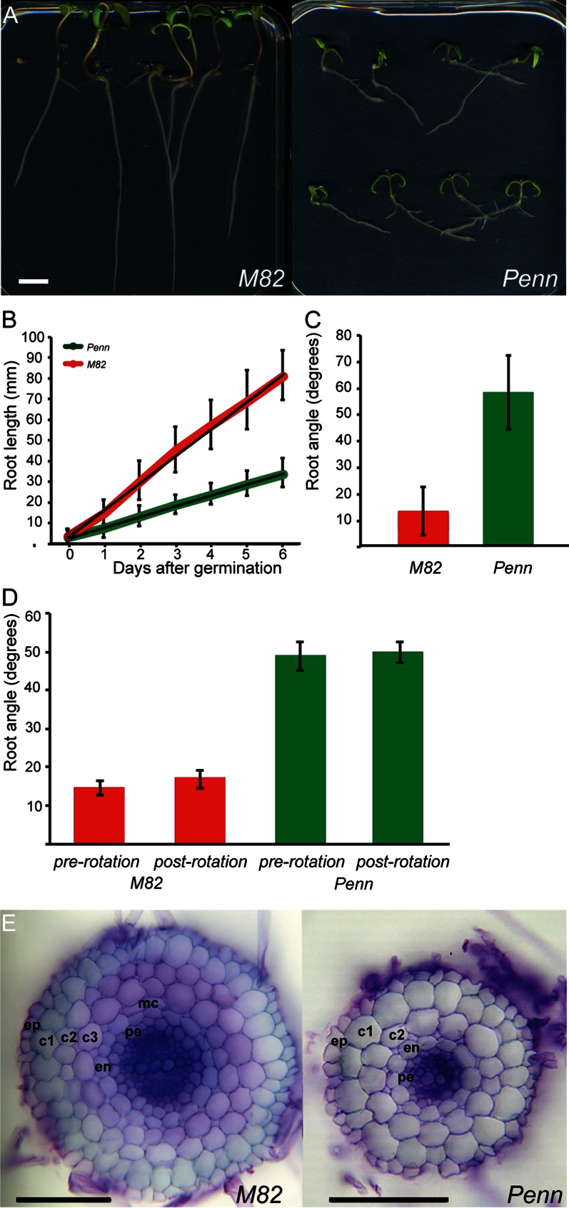
Figure 1.
Root morphology and cellular anatomy of S. lycopersicum ‘M82’ and S. pennellii (Penn). In all cases, error bars represent sd. A, cv M82 and S. pennellii root length and growth angle. Bar = 1 cm. B, Root length. All points at and following 1 d post germination are significant at P < 0.001; n = 24–60 (cv M82), n = 73–79 (S. pennellii). C, Root growth angle. The angle is significantly different at P < 0.001; n = 59 (cv M82), n = 76 (S. pennellii). D, Mean root angles before (pre) and after (post) turning growing roots 90°. Angles are significantly different between genotypes (P < 0.001) but not between treatments (P = 0.14 and 0.08 for cv M82 and S. pennellii, respectively); n = 13 (cv M82), n = 7 (S. pennellii). E, Cross sections of cv M82 and S. pennellii roots. Bar = 100 µm. Ep, Epidermis; c1, outer cortex layer; c2, S cortex layer; c3, inner cortex layer; mc, middle cortex; en, endodermis; pe, pericycle. S. pennellii is missing the c3 layer.

