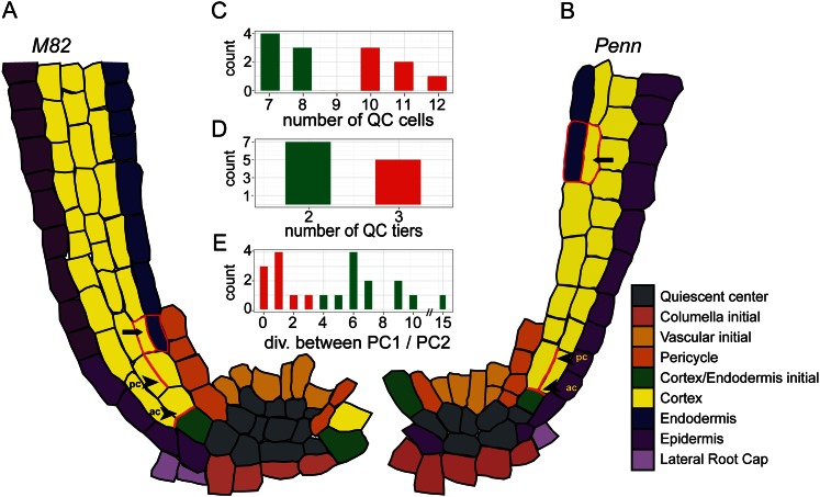
Figure 3.
Characterization of the stem cell niche of cv M82 and S. pennellii (Penn). A–B, Cellular architecture of cv M82 (A) and S. pennellii (B) stem cell niche as seen in a representative median longitudinal optical section. Pc, Periclinal division; ac, anticlinal division. C–D, Number of QC cells (C) and QC tiers (D) of cv M82 (red) and S. pennellii (green). The difference in QC cell number was significant at P = 2.48e–5 using a Student’s t test. E, The number of anticlinal divisions between the first periclinal division and the second periclinal division in cv M82 (red) and S. pennellii (green). The last periclinal division in cv M82 and S. pennellii that gives rise to the endodermis is indicated with an arrow. PC1, Periclinal division 1; PC2, periclinal division 2.

