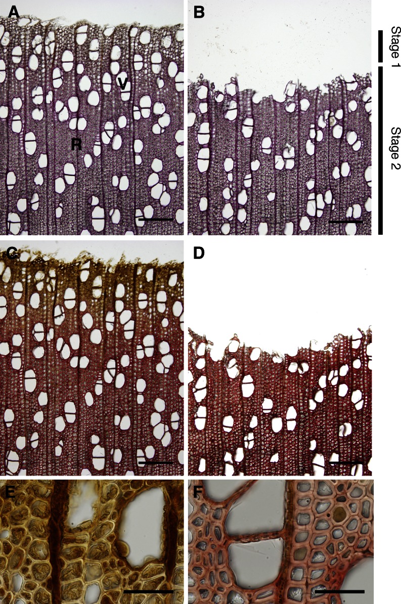
Figure 1.
Histochemical analyses of the extent of lignification of hybrid poplar. After bark removal, differentiating xylem was scraped and gathered. Images in A, C, E, and F were taken before scraping the differentiating xylem, and those in B and D were taken after scraping. A and B, Wiesner reaction of sections showing lignin distribution as red-purple staining. C to F, Mäule reaction of sections showing syringyl lignin distribution as red-purple staining. E and F show scraped xylem magnified images of C, with differentiating before lignifying (E) and lignifying vigorously in the secondary wall (F). Stage 1 contained fibers under thickening of the secondary wall; stage 2 consisted of fibers after secondary wall thickening. R, Ray parenchyma; V, vessel. Bars = 200 µm (A–D) and 50 µm (E and F).

