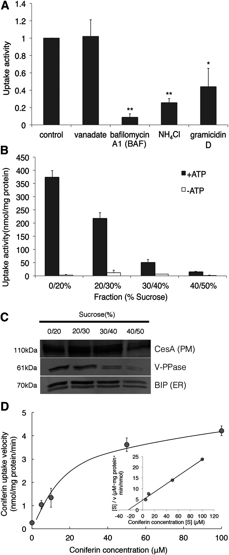
Figure 6.
Characterization of ATP-dependent coniferin uptake in Japanese cypress. A, Microsomal fractions were incubated with 50 μm coniferin and 5 mm Mg/ATP, to which vanadate (1 mm), bafilomycin A1 (BAF; 1 µm), NH4Cl (10 mm), or gramicidin D (50 µm) was added. Data are means ± sd of three replicates. *P < 0.05, **P < 0.01 compared with the control by Student’s t test. B and C, Discontinuous Suc gradient fractionation of Japanese cypress microsomes and transport assay. B, Coniferin uptake activity of each membrane fraction collected from the interface between the indicated Suc concentrations. Fractions were incubated with 50 μm coniferin in the presence or absence of 5 mm Mg/ATP. Data are means ± sd of three replicates. C, Cellulose synthase A (CesA), V-PPase, and BiP were immunodetected to confirm the purity of plasma membrane vesicles, tonoplast and endomembrane vesicles, and endoplasmic reticulum (ER) membrane vesicles, respectively. D, Coniferin uptake into membrane fractions shows saturation kinetics. Membrane fractions were incubated in the presence of 5 mm Mg/ATP and each concentration of coniferin. The inset shows Hanes-Woolf plots. Data are means ± sd of three replicates.

