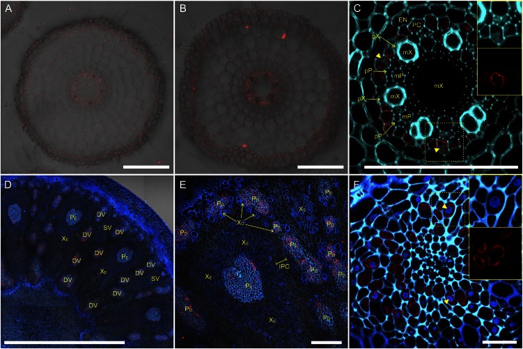
Figure 2.
Localization of OsHMA2 in rice root and node. Immunohistochemical staining of OsHMA2 with anti-OsHMA2 polyclonal antibody was performed. A and B, Root cross section at 5 mm (A) or 20 mm (B) from the apex. C, Magnified image of the stele in B. D–F, Cross section of node I at the flowering stage with different magnification. F, Close-up of phloem region of a diffuse vascular bundle. Red color indicates the OsHMA2-specific signal. Blue color indicates cell wall autofluorescence and nucleus stained by DAPI (yellow arrowheads). Insets in C and F are channel-separated magnified images at yellow-dotted areas. Bars = 100 μm (A–C and E), 1 mm (D), and 20 μm (F). Endodermis (EN), pericycle (PC), xylem vessel of proto- and metaxylem (pX and mX, respectively), and sieve tube of proto- and metaphloem (pP and mP, respectively) in the root and diffuse vascular bundle (DV), small vascular bundle (SV), xylem area of enlarged and diffuse vascular bundle (XE and XD, respectively), phloem area of enlarged and diffuse vascular bundle (PE and PD, respectively), and intervening parenchyma cells (IPC) in the node are shown.

