Figure 1.
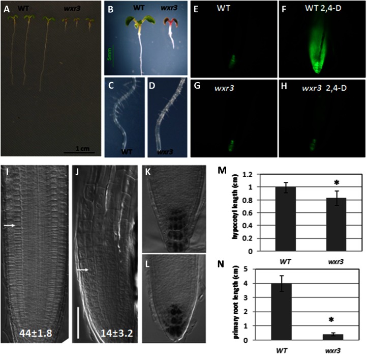
The wxr3 mutant displays severe defects in root development and 2,4-D response. A and B, Phenotypes of 10-d-old wxr3 seedlings compared with the wild type (WT). C and D, Root hair development in 10-d-old wild-type (C) and wxr3 (D) seedlings. E to H, The wxr3 mutant has dramatically reduced DR5rev:GFP level after 2,4-D treatment. Without 2,4-D, the wxr3 mutant (G) has a similar level of GFP signal to the control line (E); after 80 nm 2,4-D treatment for 12 h, the GFP level is dramatically increased in the control line (F) but not in the wxr3 mutant (H). I and J, The structure of the root apex of wild-type (I) and wxr3 (J) seedlings. The arrows denote the boundary between the meristem and the elongation zone. Numbers indicate cortical cell numbers in the meristem zone. Bar = 50 μm. K and L, Lugol staining of columella cells of wild-type (K) and wxr3 (L) roots. M, The wxr3 mutant displays a severe defect in primary root elongation compared with the wild type. N, The wxr3 mutant has a shorter hypocotyl length compared with the wild type. An ANOVA followed by Fisher’s lsd mean separation test (SPSS) were performed on the data. The asterisks indicate statistically significant differences between the wild type and the wxr3 mutant (ANOVA, P < 0.01). Error bars represent se. All seedlings in E to N were 7 d old.

