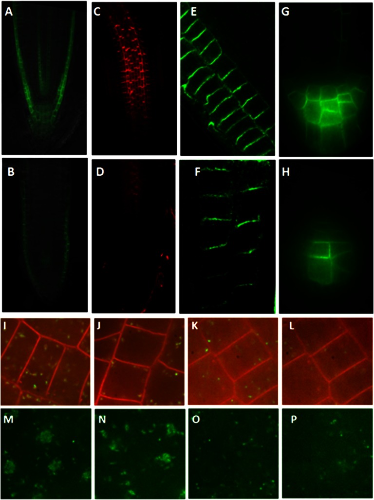
Figure 5.
The wxr3 mutant has reduced levels of auxin transporters and endosomal markers in the cell. A and B, Expression of pAUX1:AUX1-YFP in the root tip of the control line (A) and the wxr3 mutant (B). C and D, PIN1 protein levels in the root tip of the wild type (C) and the wxr3 mutant (D). E and F, PIN2 protein levels in the root tip of the wild type (E) and the wxr3 mutant (F). G and H, Expression of pPIN3:PIN3-GFP in the root tip of the control line (G) and the wxr3 mutant (H). I and J, The wxr3 mutant (J) has lower levels of VHA-a1-GFP compared with control seedlings (I). K and L, The wxr3 mutant (L) exhibits a lower level of ARA7-GFP compared with control seedlings (K). M and N, Subcellular localization of VHA-a1-GFP in the wxr3 mutant (N) and control seedlings (M) after 50 μm BFA treatment for 2 h. O and P, Subcellular localization of ARA7-GFP in the wxr3 mutant (P) and control seedlings (O) after 50 μm BFA treatment for 2 h.

