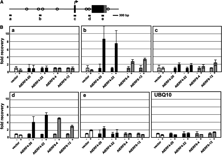
Figure 9.
Binding of AtERF4 and AtERF8 to the genomic region of ESP/ESR. A, A diagram of the genomic region of ESP/ESR. Black and gray boxes indicate the coding and untranslated regions, respectively. White circles show the position of the CCGnC motif, a target sequence of AtERF4. Thick bars below the diagram indicate the regions amplified with sets of primers in the ChIP analysis in B. B, Enrichment of the ESP/ESR sequences in the ChIP analysis. Chromatins were prepared from Pro-35S:NLS-GFP-HA (vector) and two independent lines of Pro-35S:AtERF4-HA (AtERF4) and Pro-35S:AtERF8-HA (AtERF8) Arabidopsis plants, immunoprecipitated in the absence (–) and presence (+) of antibodies, and subjected to quantitative PCR. The fold recovery was relative to the value processed in the absence of antibodies. Error bars indicate sd of technical triplicates.

