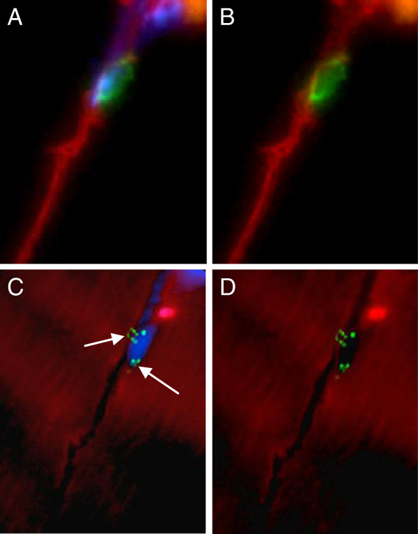
Figure 3.
Satellite cell containing two X chromosomes. Immunofluorescent staining visualized by fluorescent microscopy shows XY-FISH combined with staining for the SC marker CD56. The section was examined with a 100× objective. (A,B) CD56 (green), together with laminin-1 (red), with and without DAPI. The CD56 positive satellite cell is located just beneath the basal lamina of the muscle fiber. (C,D) The same SC after XY-FISH, with and without DAPI, showing the presence of two X chromosomes (bright green, arrows).

