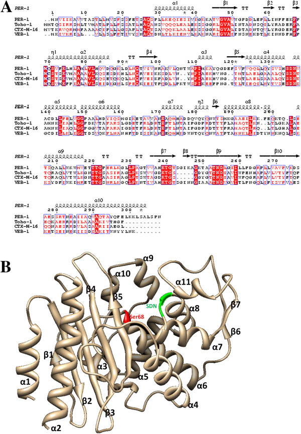
Figure 1.
Overall structure of VEB-1 and its sequence alignment with its homologue proteins. A. Sequence alignment of VEB-1 with PER-1, Toho-1 and CTX-M-16. The second structure assignment of PER-1 is labeled on the top of the sequences. B. Cartoon representation of the overall structure of VEB-1 is in light orange color. The serine active-site is colored in red, the SDN motif in green.

