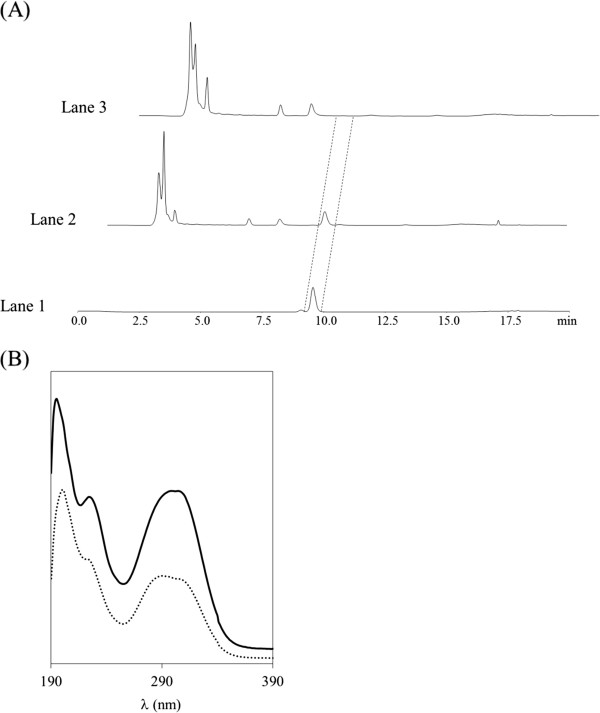
Figure 1.
Confirmation of pHCA produced by RsTAL expressing S. lividans. (A) HPLC analysis of pHCA. Lane 1: Standard sample of pHCA in acetonitrile:phosphate buffer (50 mM, pH 2.0) (20:80). Lane 2: S. lividans/pURsTAL culture supernatant. Lane 3: S. lividans/pU culture supernatant. (B) UV spectra of pHCA. Standard sample of pHCA in acetonitrile:phosphate buffer (50 mM, pH 2.0) (20:80) (solid line). The spectrum of the putative pHCA fraction isolated from the S. lividans/pURsTAL culture supernatant by HPLC is shown as a dotted line.

