
FIGURE 3. Liver histology and TUNEL staining in wild-type and jnk1 and jnk2 knock-out mice after GalN/LPS treatment.
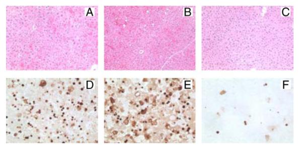
Shown are hematoxylin- and eosin-stained sections of wild-type (A), jnk1−/− (B), and jnk2−/− (C) mice at 6 h after GalN/LPS treatment. TUNEL staining at the same time point in wild-type (D), jnk1−/− (E), and jnk2−/− (F) mice (magnification 400×) is also shown.