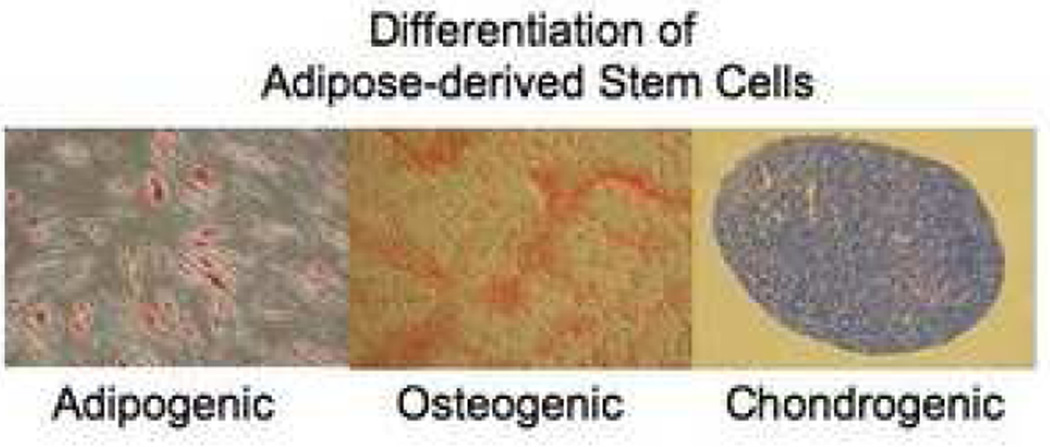
Figure 3.
Mesenchymal lineage differentiation assays for adipose-derived stem cells. Adipogenic differentiation: As the stem cells proliferate, some of these cells differentiate into preadipocytes. The preadipocytes undergo a second differentiation step and begin to fill with lipid. Lipid accumulates within the cell in small vacuoles, which appear as droplets. Osteogenic differentiation: As cells undergo osteogenic differentiation they proliferate rapidly and form tightly packed colonies. In some cases, these colonies give rise to dense nodules from which radiated highly elongated spindle-shaped cells with large nuclei. There are several methods to determine osteogenesis, such as demonstration of mineralization by staining with Alizarin red, measurement of alkaline phosphatase (AP) activity, level of calcium, and detection of lineage specific gene and protein regulations. Chondrogenic differentiation: Chondrogenesis occurs in three stages. Cartilage formation initiates when the dividing MSCs begin expressing extra cellular matrix proteins that tell them to condense into nodules. Cells in these nodules become chondrocytes and begin secreting the proteoglycans and collagen necessary for cartilage formation.

