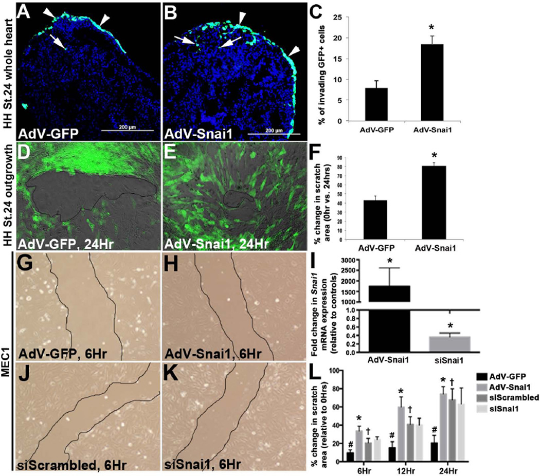
Figure 4. Snai1 enhances cell invasion from the epicardial cell layer into the myocardium.
(A-C) HH St. 24 whole heart explants were infected with AdV-GFP or AdV-Snai1 for 48 hours to label the outermost layer of the cells over the myocardium. (A, B) Tissue sections of infected whole hearts to show fate of GFP+ cells following infection with AdV-GFP (A) or GFP-tagged AdV-Snai1 (B) on the inside (arrows) and outside (arrowheads) of the developing heart. (C) Percent of invading GFP+ cells within the myocardium, over GFP+ cells on the outermost layer of the heart. (D-L) Scratch-healing assay of epicardial cell outgrowths from HH St. 24 whole heart cultures (D-F) and MEC1 cells (G-L) infected with AdV-GFP (D, G), AdV-Snai1 (E, H), siScrambled siRNA (J) or siSnai1 (K). (F) Percent change in scratch area in HH St.24 epicardial cell outgrowths following AdV treatments at 24 hours, compared to 0 hours (n=3, * =p<0.05). (I) qPCR of Snai1 expression in MEC1 cells following AdV-Snai1 and siSnai1 treatments compared to AdV-GFP and siScrambled controls respectively (n=3, * =p<0.05). (L) Percent change in scratch area in MEC1 cells following AdV-GFP, AdV-Snai1, siScrambled and siSnai1 treatments at 6, 12 and 24 hours, compared to 0 hours. #=p<0.05 AdV-GFP infections at 6, 12, and 24 hours compared to 0 hours, *=p<0.05 AdV-Snai1 at 6, 12 and 24 hours compared to AdV-GFP, †=p<0.05 siScrambled at 6, 12, and 24 hours compared to 0 hours.

