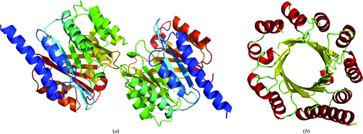
Figure 2.
X-ray crystal structure of PatF. (a) ‘Chainbows’ representation of the asymmetric unit showing the presence of two molecules. (b) Secondary-structure representation of the PatF monomer; α-helices are shown in red and β-sheets are shown in yellow. PatF is formed by a 12-stranded antiparallel β-barrel surrounded on the outside by 12 α-helices, with α-helix 1 protruding out from the rest of the structure.

