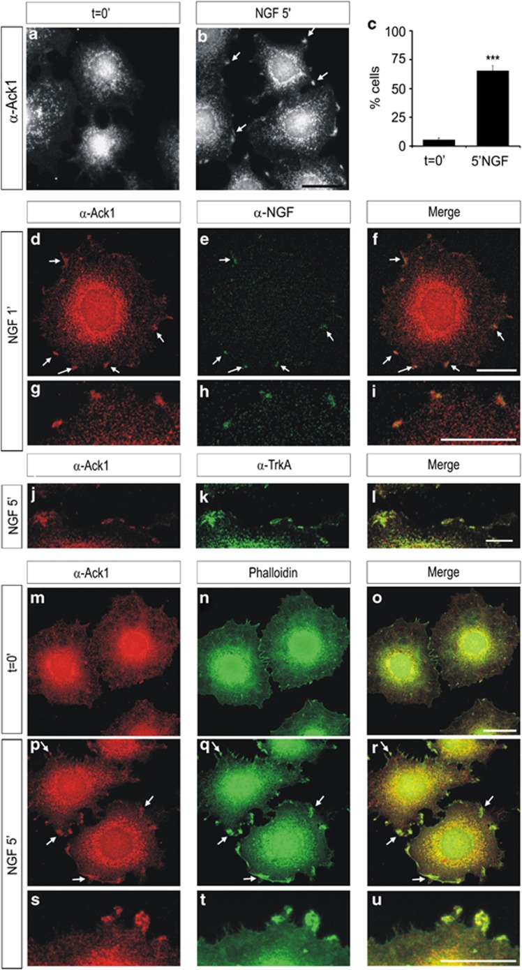
Figure 3.
Neurotrophin treatment causes a re-organization of Ack1 in PC12 cells. Immunostaining analyses of PC12 cells showing the distribution of Ack1 in untreated cells (a) and after NGF treatments (b). NGF stimuli promoted a redistribution of Ack1 protein, which is reflected in the presence of Ack1 in the cell edge (arrows). This re-organization of Ack1 was detectable in >60% of the cells (c). (d–i) Co-localization between Ack1 and NGF in PC12 cells after short stimulations with NGF. PC12 cells were incubated with anti-Ack1 and anti-NGF. As previously described, a significant pool of Ack1 protein was detected in the cell membrane and these Ack1 aggregates greatly co-localized with NGF. (j–l) Co-localization between Ack1 and TrkA in PC12 cells after a 5-min stimulation with NGF. As previously shown, a significant amount of Ack1 protein was co-localized with TrkA in cell membrane clusters. Also, Ack1 significantly co-localized with F-actin in these discrete membrane edge regions (m–u). Scale bars: 25 μm (a and b), 20 μm (d–f and m–r), and 10 μm (g–l and s–u)

