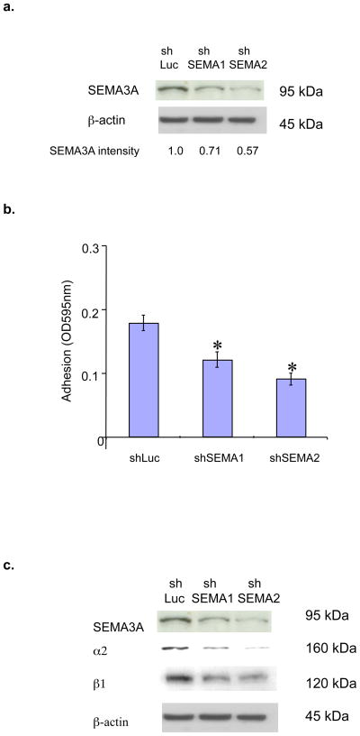
Figure 3. Endogenous SEMA3A in breast tumor cells promotes α2β1 expression and collagen adhesion.
(a) MDA-MB-231 cells were infected with lentiviruses expressing shSEMA3A RNA#1 (shSema1), shSEMA3A RNA#2 (shSema2) or shLuciferase RNA (shLuc). Equivalent amounts of protein extracted from these cells (90% confluence) were subjected to SDS-PAGE, transferred to nitrocellulose and immunoblotted with a SEMA3A or β-actin antibody, followed by the appropriate secondary antibody. The intensity of SEMA3A bands was quantified by densitometry using ImageJ software (NIH). (b) MDA-MB-231 subclones expressing shSEMA3A RNAs (shSema1 and shSema2) or shLuc RNA were grown to confluence, resuspended in serum-free medium, and plated on 96-well microtitre plates coated with bovine collagen type I (20ug/mL). After 40 min, collagen adhesion was measured as described in (A). Data are presented as the difference between the mean OD595 on BSA (from triplicate wells) and the mean OD595 on collagen (from triplicate wells) (± SD). *p < 0.05 in a student’s t-test. Similar results were obtained in two independent experiments. (c) Total cellular proteins were extracted from MDA-MB-231 subclones expressing shSEMA3A RNAs (shSema1 and shSema2) or shLuciferase (shLuc). Equivalent amounts of protein were subjected to SDS-PAGE and immunoblotted with a SEMA3A, α2, β1, or β-actin antibody, followed by the appropriate species secondary antibody.

