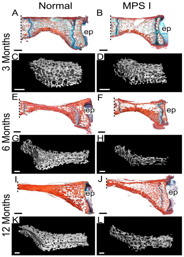
Figure 1. Histology and microCT reconstructions of C2 vertebral bone.
Mid-sagittal, histological sections, double-stained with Alcian blue (glycosaminoglycans) and picrosirius red (collagen), from normal and MPS I dogs aged 3, 6 and 12 months (A, B, E, F, I and G), with 3D microCT reconstructions of trabecular bone for the same samples (C, D, G, H, K and L). Lower trabecular bone volume is apparent for MPS I samples at all ages, but is most striking at 12 months-of-age. All histological images are oriented with the ventral (anterior) side at the bottom; ep = caudal vertebral end plate. Scale bars = 2 mm (histology) or 1 mm (microCT). Dashed lines indicate approximate location of odontoid process attachment.

