
Fig 6.
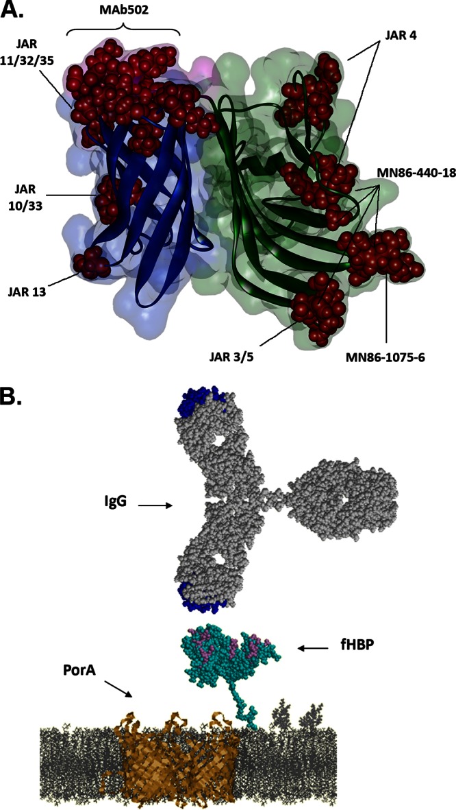
Antibody-fHBP interactions. (A) The binding residues on fHBP of bactericidal antibodies. Published bactericidal monoclonal antibody epitopes are highlighted as red spheres on the secondary structure diagram of B01 fHBP. fHBP domains are colored by domain (green, N-terminal domain; blue, C-terminal domain; pink, linker between the β-structures of two domains), with fH binding residues highlighted in purple. (B) Interaction between surface-exposed fHBP and IgG. Depicted schematically in the cell membrane are fHBP (cyan) and PorA (gold) expressed on the N. meningitidis surface; on the top, an IgG is drawn to scale. The fH binding residues on fHBP and the complementarity-determining regions on IgG are highlighted in purple and blue, respectively.