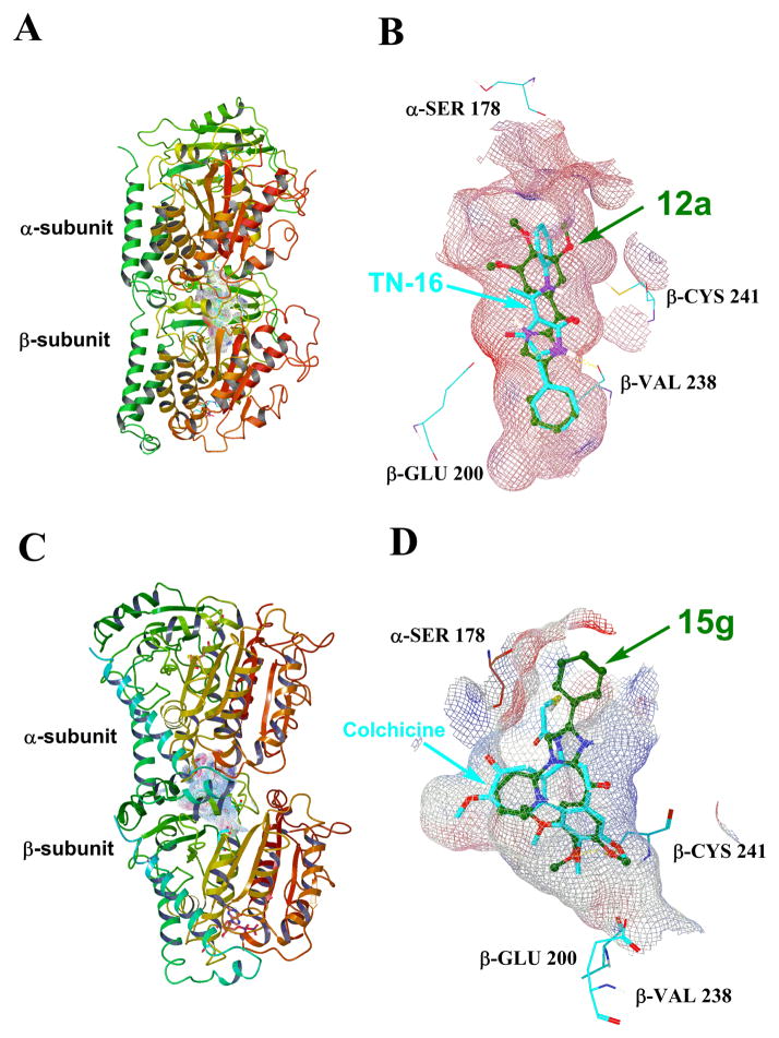
Figure 3.
(A) The overview of the binding modes of 12a and the native ligand TN-16 in tubulin crystal structure 3HKD. (B) The close view of the potential binding pose of 12a and TN-16 in 3HKD. (C) The overview of the binding modes of 15g and the native ligand colchicine in tubulin crystal structure 1SA0.(D) The close view of the potential binding pose of 15g and colchicine in 1SA0.

