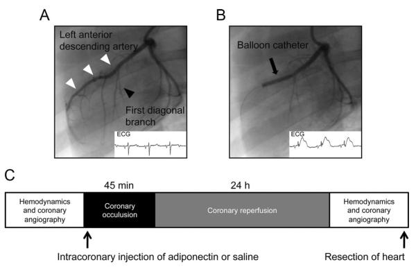
Figure 1.
Induction of myocardial I/R in pigs. A, Baseline coronary angiogram and ECG, showing the LAD (white arrows) and the first diagonal branch (black arrow). B, Coronary angiogram and ECG during the procedure, with the inflated balloon in the LAD distal to the first diagonal branch (black arrow). C, Schematic illustration of the experimental protocol.

