FIGURE 4.
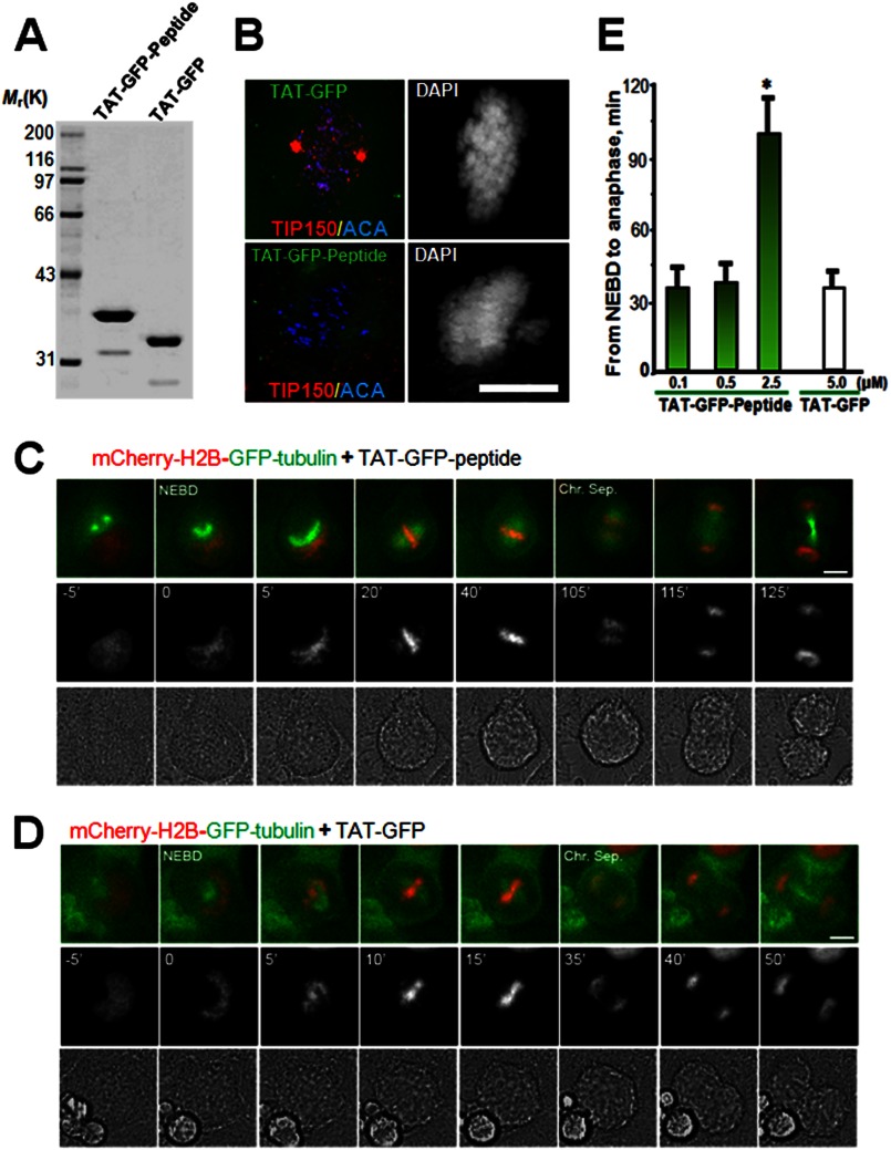
Perturbation of the EB1-TIP150 interaction prevents chromosome alignment and delays anaphase onset. A, Coomassie Blue stain SDS-polyacrylamide gel was used to assess the quality and quantities of the purified TAT-GFP-His6-tagged proteins. Bacteria expressed TAT-GFP-peptide and TAT-GFP-H6 (control) are purified with nickel-nitrilotriacetic acid affinity chromatography and desalted into DMEM. Protein concentration was determined in Bradford assay. B, TAT-GFP-peptide recombinant protein disrupts EB1-TIP150 association. Aliquots of TAT-GFP or TAT-GFP-peptide (2.5 μm) was added into culture HeLa cells for 30 min followed by fixation and examination. Note that incubation of TAT-GFP-peptide librated TIP150 from kinetochore localization and resulted in a phenotype of mitotic arrest with chromosome aberrantly aligned. C and D, HeLa cells expressing mCherry-H2B and enhanced GFP-α-tubulin were synchronized with thymidine and released for 8 h to reach prometaphase. Cells were cultured in DMEM with 500 nm, 1 or 2.5 μm TAT-GFP-TIP150-d-H6 (C) or TAT-GFP-His6 (D) at 37 °C for 1–2 h before images collection. Live cell observation and imaging were performed every 5 min. Representative images are marked especially at the time of NEBD and the time of sister chromosome separation (Chr. Sep.). Bar, 10 μm. E, quantitative analysis of the timing of cell division from NEBD to anaphase onset, which is delayed in cells treated with TAT-GFP-TIP150-d (2.5 μm). The TAT-GFP-TIP150 peptide causes mitotic delay in a dose-dependent manner. (*, p < 0.001, prometa. versus metaphase or anaphase).

