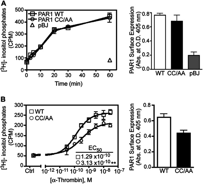
FIGURE 3.

Signaling by the palmitoylation-deficient PAR1 mutant. A, HeLa cells transfected with FLAG-PAR1 WT, CC/AA mutant, or pBJ vector were labeled with myo-[3H]inositol and stimulated with 10 nm α-thrombin for various times at 37 °C. The data (mean ± S.D.; n = 3) represent the accumulation of [3H]inositol phosphates in counts per min (cpm). Cell surface expression of PAR1 WT-, CC/AA-, and pBJ-transfected cells from the same experiment was determined by ELISA (mean ± S.D.; n = 3). B, HeLa cells transfected with FLAG-PAR1 WT or CC/AA mutant labeled with myo-[3H]inositol were treated with varying concentrations α-thrombin for 20 min at 37 °C. The data (mean ± S.D.; n = 3) represent the accumulation of [3H]inositol phosphates expressed as cpm. The EC50 values calculated for PAR1 WT (1.29 × 10⁻¹⁰ m) versus CC/AA mutant (3.13 × 10⁻¹⁰ m) were statistically significantly different (**, p < 0.01). Cell surface expression of PAR1 WT and CC/AA mutant from the same experiment was determined by ELISA (mean ± S.D.; n = 3).
