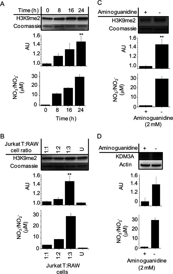
FIGURE 7.
Paracrine regulation of H3K9me2 methylation and KDM3A expression in Jurkat T cells cocultured with •NO-synthesizing RAW 264.7 cells. RAW 264.7 cells in monolayer were stimulated with LPS for 6 h. Thereafter, media were replaced with LPS-free media followed by the addition of Jurkat T cells in suspension. Nitric oxide synthesis was verified in each experiment by chemiluminescent measurements of NO3−/NO2− accumulation in the media of the cocultured cells. Immunoblot and densitometric quantifications are shown in A. Temporal changes in H3K9me2 from Jurkat T cell total histone extracts after 0–24 h coculture are shown. B, H3K9me2 measurements from Jurkat T cell total histone extracts after 24 h coculture with increasing concentrations of activated RAW 264.7 cells. U indicates incubation with nonactivated RAW 264.7 cells 1:3. C, changes in H3K9me2 from Jurkat T cell total histone extracts after 24 h coculture ± the iNOS inhibitor aminoguanidine. D, changes in KDM3A protein in whole cell lysates of Jurkat T cells after 24 h coculture ± the iNOS inhibitor aminoguanidine. All are representative immunoblots and chemiluminescent measurements of n ≥3. ** indicates p < 0.01 with respect to untreated controls, which are set arbitrarily to 1.0. AU, arbitrary unit.

